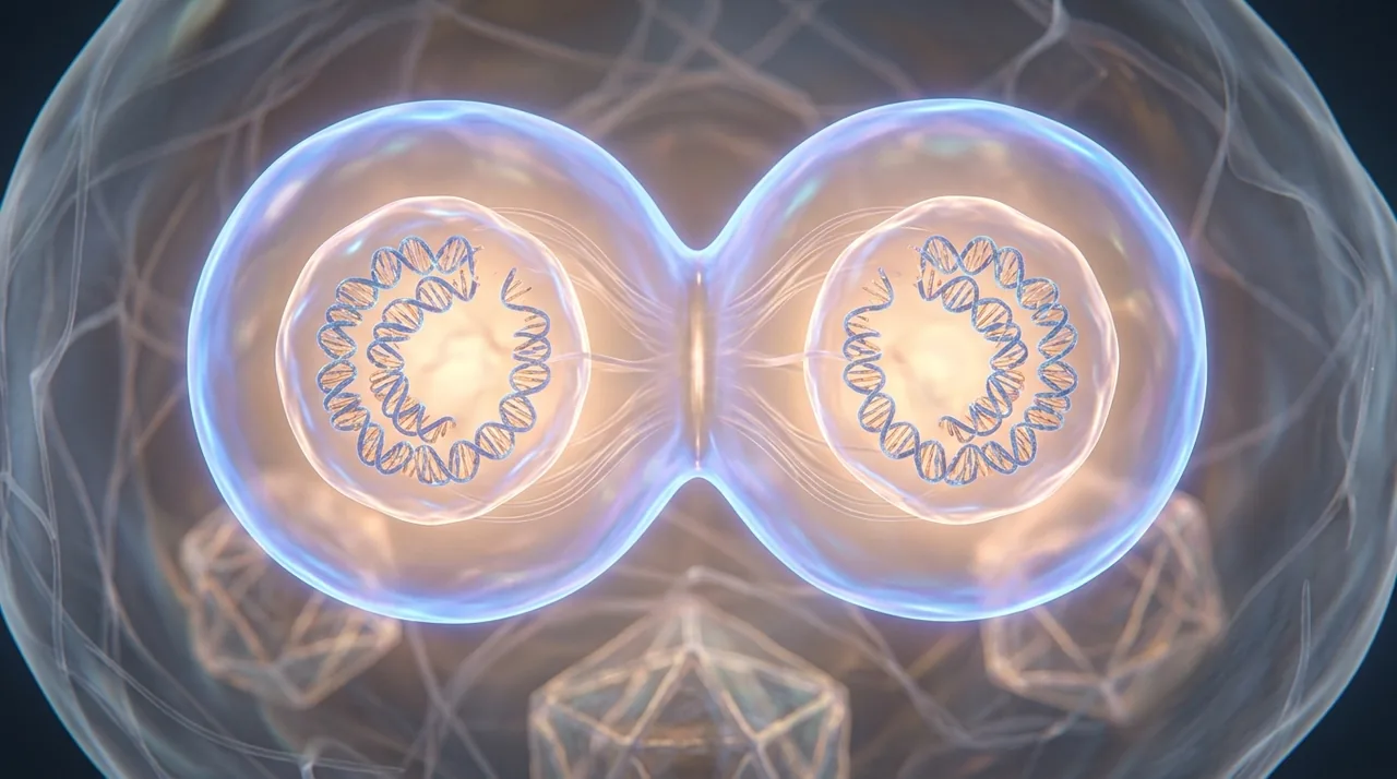
Hold your hand under a lamp. In the time it takes you to read this sentence, roughly two million of the cells in your body have split in half. The lining of your gut is replacing itself this week. The red cells in your blood have a four-month shelf life and are turned over in their millions every second. The skin you are touching right now is, by mass, mostly dead — the living version is one layer down, dividing constantly to push the dead bit off you. An adult human runs through somewhere between fifty and seventy billion cell divisions a day. None of this is conscious. None of it is supervised. And yet the error rate, when scientists go looking for it, sits at roughly one wrong letter per billion copied.
The machinery that does this is called mitosis ConceptMitosisThe process by which a eukaryotic cell divides its duplicated chromosomes evenly between two daughter nuclei. First described in detail by Walther Flemming in the 1880s, who watched salamander cells under a microscope and named the dancing threads he saw 'chromatin'. The choreography he sketched — condensation, alignment, separation, division — is still the textbook account a century and a half later.真核细胞将其复制后的染色体均等地分配到两个子核中的过程。瓦尔特·弗莱明于19世纪80年代首次对其进行了详尽描述,他在显微镜下观察蝾螈细胞,并将所见到的舞动着的丝状物命名为"染色质"。他所勾勒的那套编排——凝缩、排列、分离、分裂——在一个半世纪后的今天,仍是教科书中的标准描述。El proceso por el cual una célula eucariota divide sus cromosomas duplicados de manera equitativa entre dos núcleos hijos. Descrito en detalle por primera vez por Walther Flemming en la década de 1880, quien observó células de salamandra al microscopio y bautizó como «cromatina» los hilos danzantes que vio. La coreografía que esbozó —condensación, alineación, separación, división— sigue siendo la descripción de los libros de texto un siglo y medio después.العمليةُ التي تَقسِم بها الخليةُ حقيقيةُ النواة كروموسوماتِها المُضاعَفةَ بالتساوي بين نواتَين ابنتَين. وصفها بالتفصيل لأول مرة فالتر فليمنغ في ثمانينيات القرن التاسع عشر، إذ راقب خلايا السلمندر تحت المجهر وأطلق على الخيوط الراقصة التي رآها اسم "الكروماتين". وما تزال التراتبيةُ التي رسمها — التكاثف، والاصطفاف، والانفصال، والانقسام — هي الروايةَ المعتمدةَ في الكتب المدرسية بعد قرن ونصف.O processo pelo qual uma célula eucariótica divide seus cromossomos duplicados de maneira uniforme entre dois núcleos-filhos. Descrito pela primeira vez em detalhes por Walther Flemming na década de 1880, que observou células de salamandra ao microscópio e batizou os filamentos dançantes que viu de 'cromatina'. A coreografia que ele esboçou — condensação, alinhamento, separação, divisão — continua sendo o relato consagrado nos manuais um século e meio depois.यूकेरियोटिक कोशिका द्वारा अपने द्विगुणित गुणसूत्रों को दो संतति केंद्रकों के बीच समान रूप से विभाजित करने की प्रक्रिया। इसका विस्तृत वर्णन सर्वप्रथम 1880 के दशक में वाल्थर फ्लेमिंग ने किया, जिन्होंने सूक्ष्मदर्शी से सैलामैंडर कोशिकाओं का अवलोकन किया और जिन नर्तक तंतुओं को उन्होंने देखा, उन्हें 'क्रोमैटिन' नाम दिया। उन्होंने जिस क्रम — संघनन, संरेखण, पृथक्करण, विभाजन — का चित्रण किया, वही डेढ़ शताब्दी बाद आज भी पाठ्यपुस्तकों का प्रामाणिक विवरण है।Proses pembelahan sel eukariotik yang membagi kromosom hasil duplikasi secara merata ke dalam dua inti anak. Pertama kali dideskripsikan secara terperinci oleh Walther Flemming pada tahun 1880-an, yang mengamati sel-sel salamander di bawah mikroskop dan menamai benang-benang yang menari yang ia lihat sebagai 'kromatin'. Koreografi yang ia sketsa — kondensasi, penjajaran, pemisahan, pembelahan — masih menjadi penjelasan baku dalam buku teks satu setengah abad kemudian.Processus par lequel une cellule eucaryote répartit équitablement ses chromosomes dupliqués entre deux noyaux fils. Décrit en détail pour la première fois par Walther Flemming dans les années 1880, qui observa au microscope des cellules de salamandre et nomma « chromatine » les filaments dansants qu'il y vit. La chorégraphie qu'il esquissa — condensation, alignement, séparation, division — demeure, un siècle et demi plus tard, l'exposé des manuels.真核細胞が複製した染色体を二つの娘核へ均等に分配する過程。1880年代にヴァルター・フレミングが初めて詳細に記述したもので、彼は顕微鏡下でサンショウウオの細胞を観察し、踊るように見える糸状構造を「クロマチン」と名付けた。彼が描き出した一連の流れ——凝縮、整列、分離、分裂——は、一世紀半を経た今なお教科書的記述として通用している。Процесс, в ходе которого эукариотическая клетка равномерно распределяет удвоенные хромосомы между двумя дочерними ядрами. Впервые детально описан Вальтером Флеммингом в 1880-х годах, который наблюдал клетки саламандры под микроскопом и назвал увиденные им пляшущие нити «хроматином». Зарисованная им хореография — конденсация, выстраивание, расхождение, разделение — и по сей день, полтора столетия спустя, остаётся каноническим изложением в учебниках.Der Vorgang, durch den eine eukaryotische Zelle ihre duplizierten Chromosomen gleichmäßig auf zwei Tochterkerne verteilt. Erstmals ausführlich beschrieben von Walther Flemming in den 1880er Jahren, der Salamanderzellen unter dem Mikroskop beobachtete und die tanzenden Fäden, die er sah, „Chromatin" nannte. Die von ihm skizzierte Choreographie – Kondensation, Anordnung, Trennung, Teilung – ist anderthalb Jahrhunderte später noch immer die Lehrbuchdarstellung.진핵세포가 복제된 염색체를 두 개의 딸핵으로 균등하게 나누는 과정. 1880년대에 발터 플레밍이 현미경으로 도롱뇽 세포를 관찰하며 처음으로 상세히 기술하였고, 그가 본 춤추는 실 같은 구조에 '염색질'이라는 이름을 붙였다. 그가 묘사한 응축, 정렬, 분리, 분할의 안무는 한 세기 반이 지난 지금도 교과서적 설명으로 남아 있다., and the version playing out inside you was already old when the first animal crawled out of the sea. Single-celled eukaryotes were doing it more or less the same way 1.5 billion years ago. Everything since — the trilobites, the dinosaurs, the oak in your garden, you — is a variation on the same trick.

Two metres in a thimble
A human cell is about twenty microns across, smaller than the width of a hair. Inside it, packed into a nucleus a tenth that size, is roughly two metres of DNA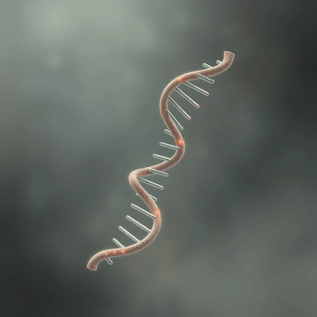 ConceptDNADeoxyribonucleic acid, the long polymer that carries genetic information in nearly every living thing. Its backbone is a chain of covalently bonded sugars and phosphates; its two strands are held to each other by hydrogen bonds between paired bases. Strong enough to copy faithfully across generations, weak enough that an enzyme can unzip the helix when a cell needs to read it.脱氧核糖核酸,是一种长链聚合物,几乎所有生物体的遗传信息均由其携带。其骨架是由糖与磷酸通过共价键连接而成的链;两条链之间则依靠配对碱基间的氢键相互结合。这种结合足够牢固,足以使遗传信息在世代间忠实地复制;又足够脆弱,使酶在细胞需要读取信息时能够将双螺旋解开。Ácido desoxirribonucleico, el largo polímero que transporta la información genética en casi todos los seres vivos. Su cadena principal es una sucesión de azúcares y fosfatos unidos mediante enlaces covalentes; sus dos hebras se mantienen unidas entre sí por puentes de hidrógeno entre bases apareadas. Suficientemente estable para copiarse fielmente a lo largo de generaciones, suficientemente lábil para que una enzima pueda desenrollar la hélice cuando la célula necesita leerla.الحمض النووي الريبوزي منقوص الأكسجين، البوليمر الطويل الذي يحمل المعلومات الوراثية في تقريباً كل كائن حي. يتألف عموده الفقري من سلسلة سكريات وفوسفات مترابطة بروابط تساهمية؛ وتتماسك سلسلتاه ببعضهما عبر روابط هيدروجينية بين القواعد النيتروجينية المتزاوجة. متين بما يكفي للنسخ الأمين عبر الأجيال، لين بما يكفي لأن تفك إنزيمٌ ضفيرتَه حين تحتاج الخلية إلى قراءته.Ácido desoxirribonucleico, o longo polímero que carrega a informação genética em quase todos os seres vivos. Sua espinha dorsal é uma cadeia de açúcares e fosfatos unidos por ligações covalentes; suas duas fitas são mantidas unidas por ligações de hidrogênio entre bases pareadas. Resistente o suficiente para ser copiado fielmente ao longo das gerações, frágil o suficiente para que uma enzima possa deszipar a hélice quando a célula precisa lê-la.डीऑक्सीराइबोन्यूक्लिक अम्ल, वह दीर्घ बहुलक जो लगभग समस्त जीवधारियों में आनुवंशिक सूचना वहन करता है। इसकी रीढ़ सहसंयोजी बंधों से जुड़ी शर्करा और फॉस्फेट की एक शृंखला है; इसके दो सूत्र युग्मित क्षारों के बीच हाइड्रोजन बंधों द्वारा परस्पर बँधे रहते हैं। इतना दृढ़ कि पीढ़ी-दर-पीढ़ी विश्वस्त रूप से प्रतिलिपि हो सके, और इतना सुनम्य कि जब कोशिका को इसे पढ़ना हो तो एक एंज़ाइम द्विकुंडली की चेन खोल सके।Asam deoksiribonukleat, polimer panjang yang membawa informasi genetik pada hampir semua makhluk hidup. Tulang punggungnya adalah rantai gula dan fosfat yang terikat secara kovalen; kedua untainya saling terhubung melalui ikatan hidrogen antara pasangan basa. Cukup kuat untuk disalin secara akurat lintas generasi, namun cukup lemah sehingga enzim dapat membuka ritsleting heliks ketika sel perlu membacanya.Acide désoxyribonucléique, le long polymère qui porte l'information génétique chez presque tous les êtres vivants. Son squelette est une chaîne de sucres et de phosphates unis par des liaisons covalentes ; ses deux brins sont maintenus l'un à l'autre par des liaisons hydrogène entre des bases appariées. Assez robuste pour se copier fidèlement d'une génération à l'autre, assez fragile pour qu'une enzyme puisse dézipper la double hélice lorsque la cellule doit la lire.デオキシリボ核酸は、ほぼすべての生物において遺伝情報を担う長鎖重合体である。その骨格は共有結合で連結された糖とリン酸の鎖から成り、二本の鎖は塩基対間の水素結合によって互いに結合されている。世代を超えて忠実に複製されるに足る強固さを備える一方、細胞がその情報を読み取る際には酵素が二重らせんを解鎖できるだけの弱さも併せ持つ。Дезоксирибонуклеиновая кислота — длинный полимер, несущий генетическую информацию практически во всех живых организмах. Остов молекулы представляет собой цепочку ковалентно связанных сахаров и фосфатных групп; две цепи удерживаются вместе водородными связями между комплементарными основаниями. Молекула достаточно стабильна, чтобы точно копироваться из поколения в поколение, и достаточно лабильна, чтобы фермент мог расплести спираль, когда клетке необходимо считать заключённую в ней информацию.Desoxyribonukleinsäure, das lange Polymer, das in nahezu allen Lebewesen die genetische Information trägt. Ihr Rückgrat ist eine Kette kovalent gebundener Zucker und Phosphate; ihre beiden Stränge werden durch Wasserstoffbrückenbindungen zwischen gepaarten Basen zusammengehalten. Stabil genug, um die Information über Generationen hinweg zuverlässig zu kopieren, und zugleich schwach genug, dass ein Enzym die Doppelhelix entfalten kann, wenn eine Zelle sie ablesen muss.디옥시리보핵산(DNA)은 거의 모든 생물에서 유전 정보를 담는 긴 중합체이다. 골격은 공유 결합으로 연결된 당과 인산의 사슬로 이루어지며, 두 가닥은 쌍을 이루는 염기 사이의 수소 결합으로 서로 결합되어 있다. 세대를 거쳐 충실히 복제될 만큼 견고하면서도, 세포가 유전 정보를 읽어야 할 때에는 효소가 이중 나선을 풀 수 있을 만큼 유연하다. coiled around protein spools called histones
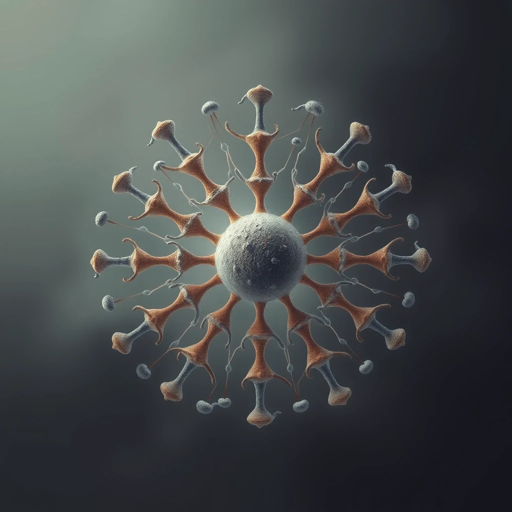 ConceptHistoneSmall, positively charged proteins around which DNA is wound to compact it inside the nucleus. Roughly 147 base pairs of DNA loop twice around an eight-histone core to form a nucleosome, the basic unit of chromatin packaging. Chemical tags added to histones — methyl groups, acetyl groups — help decide which stretches of DNA are read and which are kept silent, and are inherited through cell division.一类带正电荷的小型蛋白质,DNA 缠绕其上以便在细胞核内被压缩。约 147 个碱基对的 DNA 在由八个组蛋白构成的核心上环绕两圈,形成核小体,即染色质包装的基本单位。组蛋白上添加的化学标签——甲基、乙酰基——参与决定哪些 DNA 片段被读取、哪些被维持沉默,并可通过细胞分裂遗传下去。Pequeñas proteínas de carga positiva alrededor de las cuales se enrolla el ADN para compactarlo dentro del núcleo. Aproximadamente 147 pares de bases de ADN se enroscan dos veces alrededor de un núcleo de ocho histonas para formar un nucleosoma, la unidad básica del empaquetamiento de la cromatina. Las marcas químicas añadidas a las histonas —grupos metilo, grupos acetilo— ayudan a decidir qué tramos de ADN se leen y cuáles se mantienen silenciados, y se heredan a través de la división celular.بروتينات صغيرة موجبة الشحنة يلتفّ حولها الحمض النووي ليُضغط داخل النواة. يلتفّ نحو 147 زوجًا قاعديًا من الحمض النووي مرتين حول نواة مؤلفة من ثماني هستونات ليُكوّن النوكليوسوم، الوحدة الأساسية لتعبئة الكروماتين. تُسهم الواسمات الكيميائية المضافة إلى الهستونات — كمجموعات الميثيل ومجموعات الأسيتيل — في تحديد أيّ أجزاء الحمض النووي تُقرأ وأيّها يبقى صامتًا، وتُتوارَث عبر الانقسام الخلوي.Pequenas proteínas com carga positiva em torno das quais o DNA se enrola para se compactar dentro do núcleo. Aproximadamente 147 pares de bases de DNA dão duas voltas em torno de um cerne de oito histonas para formar um nucleossomo, a unidade básica do empacotamento da cromatina. Marcas químicas adicionadas às histonas — grupos metil, grupos acetil — ajudam a determinar quais trechos de DNA são lidos e quais são mantidos silenciados, e são herdadas através da divisão celular.छोटे, धनात्मक आवेश वाले प्रोटीन जिनके चारों ओर DNA लिपटकर केन्द्रक के भीतर सघन रूप धारण कर लेता है। लगभग 147 क्षार-युग्म DNA आठ हिस्टोनों के एक कोर के चारों ओर दो बार लिपटकर न्यूक्लियोसोम बनाता है, जो क्रोमैटिन पैकेजिंग की मूल इकाई है। हिस्टोनों पर जुड़े रासायनिक चिह्न — मेथिल समूह, ऐसिटिल समूह — यह तय करने में सहायक होते हैं कि DNA के कौन-से खंड पढ़े जाएँगे और कौन-से मौन रखे जाएँगे, और ये कोशिका विभाजन के माध्यम से वंशागत होते हैं।Protein kecil bermuatan positif yang menjadi penggulung DNA untuk memadatkannya di dalam inti sel. Sekitar 147 pasang basa DNA melilit dua kali pada inti delapan histon untuk membentuk nukleosom, unit dasar pengemasan kromatin. Penanda kimia yang ditambahkan pada histon — gugus metil, gugus asetil — turut menentukan ruas DNA mana yang dibaca dan mana yang dibungkam, dan diwariskan melalui pembelahan sel.Petites protéines chargées positivement autour desquelles l'ADN s'enroule pour se compacter dans le noyau. Environ 147 paires de bases d'ADN s'enroulent deux fois autour d'un cœur de huit histones pour former un nucléosome, l'unité de base du compactage de la chromatine. Des marques chimiques ajoutées aux histones — groupes méthyle, groupes acétyle — contribuent à déterminer quelles portions d'ADN sont lues et lesquelles sont maintenues silencieuses, et se transmettent au fil des divisions cellulaires.DNAを核内で凝縮するために巻きつけられる、小さく正に帯電したタンパク質。約147塩基対のDNAが8個のヒストンから成る中心粒の周囲を2回巻きついてヌクレオソームを形成し、これがクロマチン包装の基本単位となる。ヒストンに付加される化学的標識──メチル基やアセチル基など──は、DNAのどの領域が読み取られ、どの領域が沈黙したまま保たれるかを決定する一助となり、細胞分裂を通じて受け継がれる。Небольшие положительно заряженные белки, вокруг которых наматывается ДНК для её компактизации внутри ядра. Около 147 пар оснований ДНК дважды обвивают восьмигистоновое ядро, образуя нуклеосому — базовую единицу упаковки хроматина. Химические метки, присоединяемые к гистонам, — метильные и ацетильные группы — помогают определять, какие участки ДНК считываются, а какие остаются молчащими, и наследуются при клеточном делении.Kleine, positiv geladene Proteine, um die DNA gewickelt wird, um sie im Zellkern zu verdichten. Etwa 147 Basenpaare DNA winden sich zweimal um einen Kern aus acht Histonen und bilden so ein Nukleosom, die Grundeinheit der Chromatinverpackung. Chemische Markierungen an Histonen – Methylgruppen, Acetylgruppen – tragen dazu bei zu bestimmen, welche DNA-Abschnitte abgelesen und welche stillgelegt werden, und werden über die Zellteilung weitervererbt.DNA를 핵 안에서 압축시키기 위해 그 주위에 감기는 작고 양전하를 띤 단백질. 약 147개의 염기쌍이 8개의 히스톤 코어를 두 번 감아 뉴클레오솜, 즉 염색질 포장의 기본 단위를 형성한다. 히스톤에 부착되는 화학적 표지 — 메틸기, 아세틸기 — 는 DNA의 어떤 부분이 읽히고 어떤 부분이 침묵 상태로 유지될지 결정하는 데 관여하며, 세포 분열을 통해 유전된다. and then folded, and folded again, into 46 dense bundles. These are the chromosomes, and for most of a cell's life you cannot see them as separate things — they exist as a tangled mess called chromatin.

When a cell decides to divide, the first thing it does is copy that entire two-metre archive. An enzyme called helicase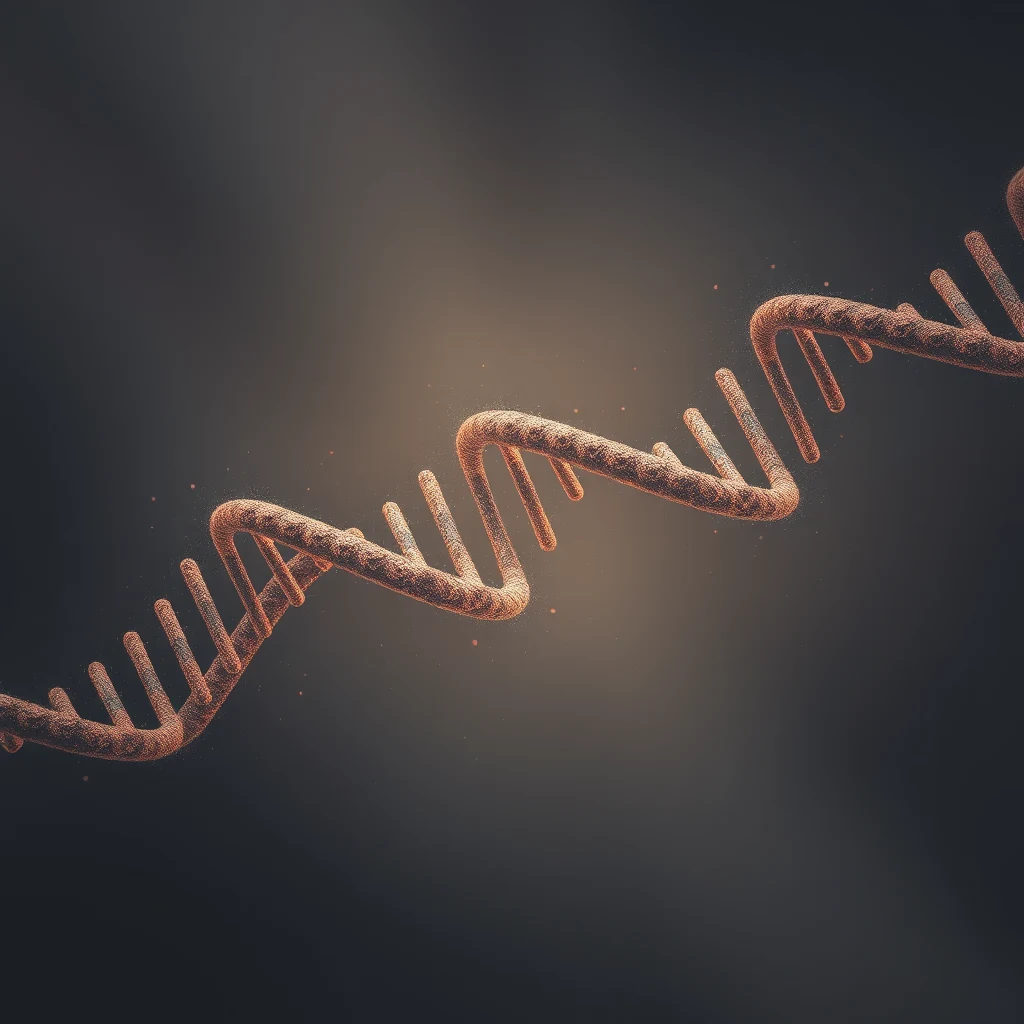 ConceptHelicaseAn enzyme that unwinds the DNA double helix ahead of replication, breaking the hydrogen bonds between paired bases and separating the two strands. Helicases burn ATP for fuel and travel along DNA at roughly a thousand base pairs per second in human cells. Without them, the polymerases that read and copy DNA would have nothing to read; the two strands are too tightly wound to access otherwise.一种在复制过程中先行解开DNA双螺旋的酶,能够断裂配对碱基之间的氢键,使两条链分离。解旋酶以ATP为燃料,在人类细胞中沿DNA以每秒约一千个碱基对的速度移动。没有它们,负责读取和复制DNA的聚合酶便无从读起;两条链缠绕得过于紧密,否则无法触及。Enzima que desenrolla la doble hélice del ADN por delante de la replicación, rompiendo los enlaces de hidrógeno entre las bases apareadas y separando las dos hebras. Las helicasas consumen ATP como combustible y se desplazan a lo largo del ADN a aproximadamente mil pares de bases por segundo en las células humanas. Sin ellas, las polimerasas que leen y copian el ADN no tendrían nada que leer; las dos hebras están demasiado fuertemente enrolladas para acceder a ellas de otro modo.إنزيم يفك التفاف الحلزون المزدوج للحمض النووي أمام عملية التضاعف، فيكسر الروابط الهيدروجينية بين القواعد المتزاوجة ويفصل الشريطين عن بعضهما. تستهلك إنزيمات الهليكاز جزيئات الـATP وقودًا لها، وتنتقل على طول الحمض النووي بسرعة تقارب ألف زوج من القواعد في الثانية داخل الخلايا البشرية. ولولاها لما وجدت إنزيمات البوليميراز التي تقرأ الحمض النووي وتنسخه ما تقرؤه؛ إذ يكون الشريطان ملتفّين بإحكام يحول دون الوصول إليهما بأي طريقة أخرى.Uma enzima que desenrola a dupla hélice de DNA à frente da replicação, rompendo as ligações de hidrogênio entre as bases pareadas e separando as duas fitas. As helicases queimam ATP como combustível e percorrem o DNA a aproximadamente mil pares de bases por segundo nas células humanas. Sem elas, as polimerases que leem e copiam o DNA não teriam o que ler; as duas fitas estão enroladas de forma demasiado compacta para serem acessadas de outro modo.एक एंज़ाइम जो प्रतिकृति से पहले DNA की द्विकुंडली को खोलता है, युग्मित क्षारों के बीच के हाइड्रोजन बंधों को तोड़ता है और दोनों लड़ियों को अलग करता है। हेलिकेज़ ईंधन के रूप में ATP का उपभोग करते हैं और मानव कोशिकाओं में DNA के साथ लगभग एक हज़ार क्षार-युग्म प्रति सेकंड की गति से चलते हैं। इनके बिना, DNA को पढ़ने और उसकी प्रतिलिपि बनाने वाले पॉलिमरेज़ के पास पढ़ने के लिए कुछ नहीं होता; दोनों लड़ियाँ अन्यथा इतनी कसकर लिपटी होती हैं कि उन तक पहुँचा नहीं जा सकता।Enzim yang membuka untai ganda DNA menjelang replikasi, memutus ikatan hidrogen antara basa berpasangan dan memisahkan kedua untainya. Helikase membakar ATP sebagai bahan bakar dan bergerak sepanjang DNA dengan laju sekitar seribu pasangan basa per detik pada sel manusia. Tanpanya, polimerase yang membaca dan menyalin DNA tidak akan memiliki sesuatu untuk dibaca; kedua untai tersebut terlalu rapat terpilin untuk dapat diakses dengan cara lain.Enzyme qui déroule la double hélice d'ADN en amont de la réplication, en rompant les liaisons hydrogène entre les bases appariées et en séparant les deux brins. Les hélicases consomment de l'ATP comme carburant et se déplacent le long de l'ADN à environ mille paires de bases par seconde dans les cellules humaines. Sans elles, les polymérases qui lisent et copient l'ADN n'auraient rien à lire ; les deux brins sont autrement trop étroitement enroulés pour être accessibles.DNA二重らせんを複製に先立って巻き戻し、塩基対間の水素結合を切断して二本の鎖を分離する酵素。ヘリカーゼはATPを燃料として消費し、ヒト細胞ではDNA上を毎秒およそ1,000塩基対の速度で進む。これがなければ、DNAを読み取り複製するポリメラーゼは読むべき鋳型を得られず、二本鎖はあまりに強く巻きついているため他の手段では接近できない。Фермент, расщепляющий двойную спираль ДНК перед репликацией: он разрывает водородные связи между парными основаниями и разделяет две цепи. Хеликазы расходуют АТФ в качестве топлива и движутся вдоль ДНК со скоростью около тысячи пар оснований в секунду в клетках человека. Без них полимеразам, считывающим и копирующим ДНК, было бы нечего читать: две цепи слишком плотно скручены, чтобы получить к ним доступ иным способом.Ein Enzym, das die DNA-Doppelhelix vor der Replikation entwindet, indem es die Wasserstoffbrückenbindungen zwischen den gepaarten Basen aufbricht und die beiden Stränge voneinander trennt. Helikasen verbrauchen ATP als Energiequelle und bewegen sich in menschlichen Zellen mit einer Geschwindigkeit von etwa tausend Basenpaaren pro Sekunde an der DNA entlang. Ohne sie hätten die Polymerasen, die DNA lesen und kopieren, nichts zu lesen; die beiden Stränge sind ansonsten zu eng umeinander gewunden, um zugänglich zu sein.복제에 앞서 DNA 이중나선을 풀어, 짝지어진 염기 사이의 수소 결합을 끊고 두 가닥을 분리하는 효소. 헬리케이스는 ATP를 연료로 소모하며, 인간 세포에서는 DNA를 따라 초당 약 1,000 염기쌍의 속도로 이동한다. 이들이 없다면 DNA를 읽어 복제하는 중합효소는 읽을 거리가 없어진다. 두 가닥은 그대로는 너무 단단히 감겨 있어 접근할 수 없기 때문이다. runs along the double helix prying the two strands apart like the teeth of a zipper. Behind it, DNA polymerase
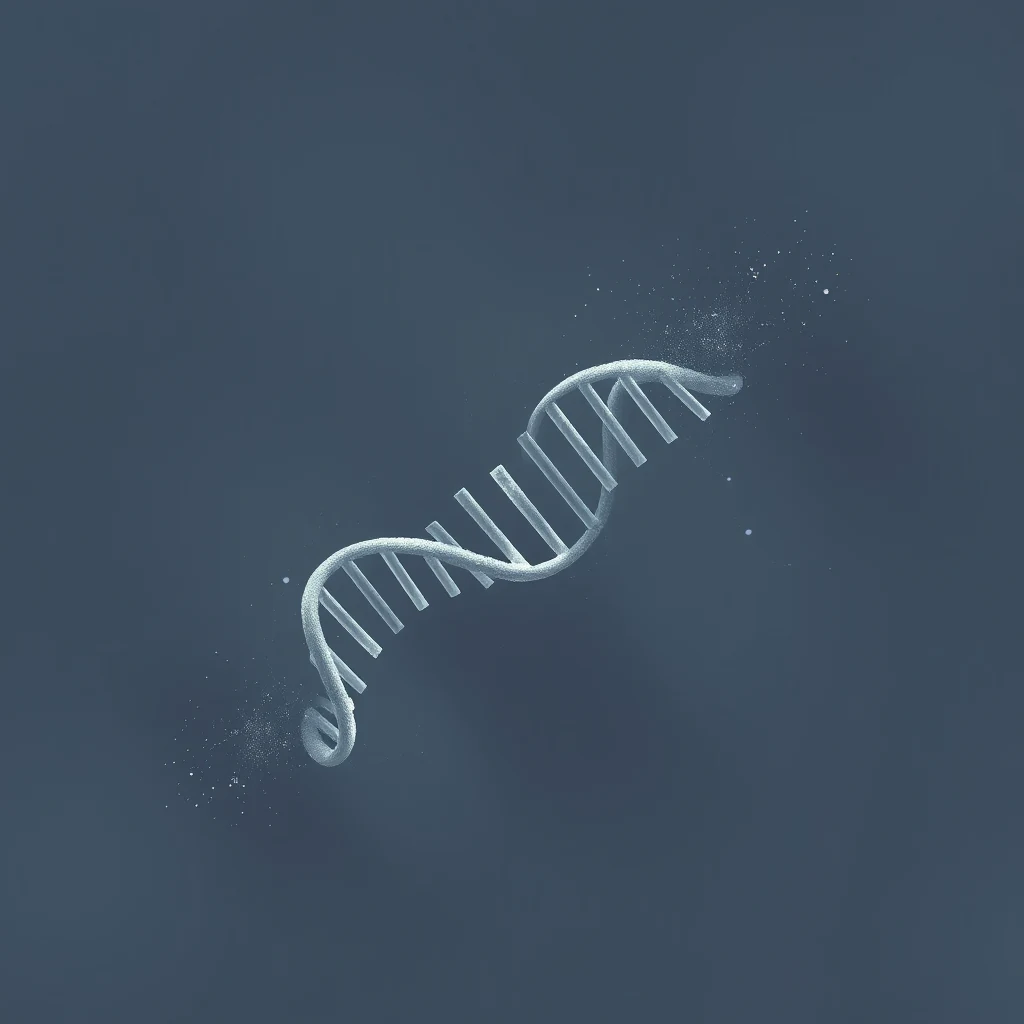 ConceptDNA polymeraseThe enzyme family that synthesises DNA by adding nucleotides to a growing strand, using a template strand as a guide. Discovered in 1956 by Arthur Kornberg, who isolated it from E. coli and won the Nobel Prize three years later. Humans have at least fifteen polymerases with specialised roles in replication, repair, and translesion synthesis.DNA聚合酶是通过模板链作为指导,将核苷酸添加到延伸链上来合成DNA的酶家族。1956年由阿瑟·科恩伯格发现,他从大肠杆菌中分离出了该酶,并于三年后获得了诺贝尔奖。人类拥有至少十五种DNA聚合酶,它们在DNA复制、修复和跨损伤合成中承担着专门的职责。Familia de enzimas que sintetiza ADN añadiendo nucleótidos a una cadena en crecimiento, usando una cadena plantilla como guía. Descubierta en 1956 por Arthur Kornberg, quien la aisló de E. coli y ganó el Nobel tres años después. Los humanos tienen al menos quince polimerasas con funciones especializadas en la replicación, reparación y síntesis a través de lesiones del ADN.عائلة الإنزيمات التي تصنع الحمض النووي عن طريق إضافة النيوكليوتيدات إلى سلسلة نامية، باستخدام سلسلة قوالب كدليل. اكتشفه آرثر كورنبرغ عام 1956، الذي عزله من الإشريكية القولونية وحصل على جائزة نوبل بعد ثلاث سنوات. يمتلك البشر ما لا يقل عن خمسة عشر إنزيماً من بوليميراز الحمض النووي ذات أدوار متخصصة في التضاعف والإصلاح.A família de enzimas que sintetiza o DNA adicionando nucleotídeos a uma fita em crescimento, usando uma fita molde como guia. Descoberta em 1956 por Arthur Kornberg, que a isolou de E. coli e ganhou o Prêmio Nobel três anos depois. Os humanos têm pelo menos quinze polimerases com papéis especializados na replicação, reparo e síntese de translesão.एंजाइम परिवार जो एक गाइड के रूप में एक टेम्पलेट स्ट्रैंड का उपयोग करके बढ़ते स्ट्रैंड में न्यूक्लियोटाइड जोड़कर डीएनए का संश्लेषण करता है, जिसे डीएनए पोलीमरेज़ (DNA polymerase) कहा जाता है। 1956 में आर्थर कॉर्नबर्ग द्वारा खोजा गया, जिन्होंने इसे ई. कोलाई से अलग किया और तीन साल बाद नोबेल पुरस्कार जीता। मनुष्यों में प्रतिकृति और मरम्मत में विशिष्ट भूमिकाओं वाले कम से कम पंद्रह पोलीमरेज़ होते हैं।DNA polimerase adalah keluarga enzim yang menyintesis DNA dengan menambahkan nukleotida ke untai yang sedang tumbuh, dipandu oleh untai cetakan. Ditemukan pada tahun 1956 oleh Arthur Kornberg, yang mengisolasinya dari E. coli dan memenangkan Hadiah Nobel tiga tahun kemudian. Manusia memiliki setidaknya lima belas polimerase dengan peran khusus dalam replikasi, perbaikan, dan sintesis translesi.La famille d'enzymes qui synthétise l'ADN en ajoutant des nucléotides à un brin en croissance, en utilisant un brin matrice comme guide. Découverte en 1956 par Arthur Kornberg, qui l'a isolée d'E. coli et a obtenu le prix Nobel trois ans plus tard. L'homme possède au moins quinze polymérases aux rôles spécialisés dans la réplication, la réparation et la synthèse translésionnelle.DNAポリメラーゼは、鋳型となるDNA鎖をガイドとして、伸長中の鎖にヌクレオチドを追加することでDNAを合成する酵素群である。1956年にアーサー・コーンバーグによって大腸菌から初めて単離され、彼はその3年後にノーベル賞を受賞した。ヒトは、DNAの複製、修復、および損傷乗り越え合成においてそれぞれ特異的な役割を担う少なくとも15種類のポリメラーゼを持っている。Семейство ферментов, синтезирующих ДНК путем добавления нуклеотидов к растущей цепи с использованием матричной цепи в качестве шаблона. Открыт в 1956 году Артуром Корнбергом, который выделил его из E. coli и через три года получил Нобелевскую премию. У человека имеется не менее пятнадцати полимераз со специализированными функциями в репликации и репарации ДНК.Die Enzymfamilie, die DNA synthetisiert, indem sie Nukleotide an einen wachsenden Strang anfügt, wobei ein Matrizenstrang als Vorlage dient. 1956 von Arthur Kornberg entdeckt, der sie aus E. coli isolierte und drei Jahre später den Nobelpreis erhielt. Menschen besitzen mindestens fünfzehn Polymerasen mit spezialisierten Aufgaben bei der Replikation, Reparatur und Translesionssynthese.DNA 중합효소(DNA polymerase)는 주형 가닥을 바탕으로 새로 합성되는 가닥에 뉴클레오타이드를 추가하여 DNA를 합성하는 효소 제품군이다. 1956년 아서 콘버그가 대장균에서 최초로 단리하여 3년 후 노벨상을 수상했다. 인간은 복제, 손상 복구, 손상 통과 합성(translesion synthesis) 등에서 각기 다른 특화된 역할을 수행하는 최소 15가지 종류의 중합효소를 보유하고 있다. reads each exposed strand and builds the complementary partner, base by base, at a rate of around fifty nucleotides per second. There are roughly three billion base pairs to get through. To finish in eight hours, the cell starts copying from thousands of points along the DNA at once.

The accuracy is not free. Polymerase itself gets about one base in 100,000 wrong. A built-in proofreading function — the enzyme reverses, snips out the mistake, and tries again — pushes that to one in ten million. A second system called mismatch repair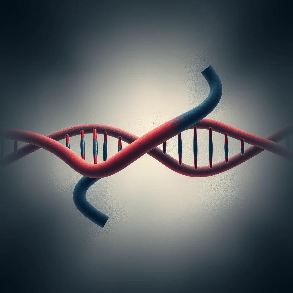 ConceptMismatch repairA genome surveillance system that scans newly replicated DNA for base-pair mismatches the polymerase missed. Proteins of the MutS and MutL families recognise the distortion, excise a stretch of the new strand, and call in polymerase to refill it. Inherited defects in human mismatch repair cause Lynch syndrome, a hereditary predisposition to colorectal and other cancers.错配修复是一种基因组监视系统,用于扫描新复制的DNA,寻找聚合酶遗漏的碱基对错配。MutS和MutL家族的蛋白质能识别这种螺旋畸变,切除新合成链上的一段,并召集聚合酶重新填补空缺。人类错配修复基因的遗传缺陷会导致林奇综合征,这是一种对结直肠癌及其他癌症的遗传易感性疾病。El sistema de reparación de apareamientos erróneos vigila el genoma escaneando el ADN recién replicado para detectar fallos que la polimerasa pasó por alto. Proteínas de las familias MutS y MutL reconocen la distorción, extirpan un tramo de la nueva cadena y reclutan polimerasa para rellenarlo. Los defectos heredados causan el síndrome de Lynch, predisposición al cáncer colorrectal.إصلاح عدم التطابق هو نظام مراقبة الجينوم الذي يفحص الحمض النووي المتضاعف حديثاً بحثاً عن أخطاء اقتران القواعد التي أغفلها البوليميراز. تتعرف بروتينات عائلتي (MutS) و (MutL) على التشويه، وتستأصل جزءاً من السلسلة الجديدة، وتستدعي البوليميراز لإعادة تعبئته. وتسبب العيوب الموروثة في هذا النظام متلازمة لينش، وهي استعداد وراثي للسرطان.O reparo de incompatibilidade de bases é um sistema de vigilância genômica que varre o DNA recém-replicado em busca de pareamentos errôneos que a polimerase perdeu. Proteínas das famílias MutS e MutL reconhecem a distorção, excisam um trecho da nova fita e recrutam a polimerase para preenchê-lo. Defeitos herdados no reparo causam a síndrome de Lynch, uma predisposição ao câncer.मिसमैच रिपेयर (Mismatch repair) एक जीनोम निगरानी प्रणाली है जो पोलीमरेज़ द्वारा छूटे गए बेस-पेयर बेमेल के लिए नव प्रतिकृति डीएनए को स्कैन करती है। MutS और MutL परिवारों के प्रोटीन विरूपण को पहचानते हैं, नए स्ट्रैंड के एक हिस्से को हटाते हैं, और इसे फिर से भरने के लिए पोलीमरेज़ को बुलाते हैं। विरासत में मिले दोष लिंच सिंड्रोम का कारण बनते हैं।Perbaikan salah pasang adalah sistem pengawasan genom yang memindai DNA yang baru direplikasi untuk mencari salah pasang basa yang terlewat oleh polimerase. Protein dari famili MutS dan MutL mengenali distorsi tersebut, memotong bagian untai baru, lalu memanggil polimerase untuk mengisinya kembali. Kerusakan genetik pada sistem ini menyebabkan sindrom Lynch.La réparation des mésappariements est un système de surveillance du génome qui scanne l'ADN néosynthétisé à la recherche des erreurs de copie échappées à la polymérase. Les protéines MutS et MutL repèrent la distorsion, éliminent un segment du nouveau brin et recrutent une polymérase pour combler la brèche. Les défauts hérités de ce système causent le syndrome de Lynch.ミスマッチ修修復は、新しく複製されたDNAを走査し、DNAポリメラーゼが看過した塩基対のミスマッチを検出するゲノム監視システムである。MutSおよびMutLファミリーのタンパク質が構造の歪みを認識し、新生鎖の異常部位を切り取り、ポリメラーゼを動員して再合成を行う。ヒトのミスマッチ修復遺伝子の遗传的欠損は、大腸癌などの遺伝性素因であるリンチ症候群を引き起こす。Репарация несоответствия — это система надзора за геномом, которая сканирует новосинтезированную ДНК на предмет неспаренных оснований, пропущенных полимеразой. Белки семейств MutS и MutL распознают искажение, вырезают участок новой цепи и привлекают полимеразу для его восстановления. Наследственные дефекты репарации у человека вызывают синдром Линча.Die Mismatch-Reparatur ist ein Überwachungssystem des Genoms, das neu replizierte DNA auf Basenfehlpaarungen scannt, die der Polymerase entgangen sind. Proteine der MutS- und MutL-Familien erkennen die Verzerrung, schneiden ein Stück des neuen Strangs heraus und rufen Polymerase zum Auffüllen herbei. Vererbte Defekte beim Menschen verursachen das Lynch-Syndrom.미스매치 복구(mismatch repair)는 새로 복제된 DNA 가닥을 탐색하여 중합효소가 놓치고 지나간 잘못된 염기쌍 결합을 찾아 수정하는 게놈 감시 체계이다. MutS 및 MutL 단백질 군이 나선 구조의 왜곡을 인지하여 오류가 발생한 신생 가닥의 일부를 절단해 제거하면, 중합효소가 호출되어 빈 공간을 다시 채운다. 인간 미스매치 복구 유전자의 선천적 결함은 대장암 등을 유발하는 린치 증후군의 원인이 된다. sweeps in afterwards and finds most of what is left. The final error rate, end to end, is around 10⁻⁹. Copy the King James Bible a thousand times by hand and you would be doing well to match it.
The dance
Once the DNA is doubled, the cell has 92 chromosomes where it had 46, each new chromosome attached to its original twin at a waist called the centromere. Then the choreography starts. The nuclear envelope dissolves. From two points at opposite ends of the cell, protein cables called microtubules ConceptMicrotubuleHollow protein tubes, about 25 nanometres across, built from a protein called tubulin. They form the cytoskeleton that gives a cell its shape and serve as tracks along which cargo is hauled. During mitosis they assemble into the spindle that drags chromosomes apart. Several chemotherapy drugs, including paclitaxel from the Pacific yew, work by jamming microtubule assembly and stalling cell division.中空的蛋白质管,直径约25纳米,由一种名为微管蛋白的蛋白质构成。它们组成细胞骨架,赋予细胞形状,并充当运输货物的轨道。有丝分裂期间,它们组装成纺锤体,将染色体牵拉分开。多种化疗药物,包括从太平洋紫杉中提取的紫杉醇,通过阻碍微管组装、停滞细胞分裂而发挥作用。Tubos proteicos huecos, de unos 25 nanómetros de diámetro, formados a partir de una proteína llamada tubulina. Constituyen el citoesqueleto que confiere a la célula su forma y sirven de vías por las que se transporta la carga celular. Durante la mitosis se ensamblan en el huso que separa los cromosomas. Varios fármacos quimioterápicos, entre ellos el paclitaxel del tejo del Pacífico, actúan bloqueando el ensamblaje de los microtúbulos y deteniendo la división celular.أنابيب بروتينية مجوّفة يبلغ قطرها نحو 25 نانومترًا، مبنية من بروتين يُسمى التوبيولين. تُشكّل الهيكل الخلوي الذي يمنح الخلية شكلها، وتعمل بمثابة مسارات تُنقل عبرها الحمولات. وأثناء الانقسام الفتيلي، تتجمّع لتكوّن المغزل الذي يسحب الكروموسومات بعيدًا عن بعضها. ويعمل عدد من أدوية العلاج الكيميائي، من بينها الباكليتاكسيل المستخلص من شجرة الطقسوس الباسيفيكي، عبر تعطيل تجمّع الأنابيب الدقيقة وإيقاف انقسام الخلية.Tubos proteicos ocos, com cerca de 25 nanómetros de diâmetro, formados por uma proteína chamada tubulina. Constituem o citoesqueleto que confere a forma à célula e funcionam como vias ao longo das quais é transportada a carga celular. Durante a mitose, organizam-se no fuso que separa os cromossomas. Vários fármacos quimioterápicos, incluindo o paclitaxel, extraído do teixo-do-pacífico, atuam bloqueando a montagem dos microtúbulos e suspendendo a divisão celular.लगभग 25 नैनोमीटर व्यास वाली खोखली प्रोटीन नलिकाएँ, जो ट्यूबुलिन नामक प्रोटीन से बनी होती हैं। ये कोशिकाद्रव्यपिंजर (साइटोस्केलेटन) का निर्माण करती हैं जो कोशिका को उसका आकार देता है, और ऐसे पथ का काम करती हैं जिन पर माल ढोया जाता है। समसूत्री विभाजन (माइटोसिस) के दौरान ये जुड़कर तर्कु (स्पिंडल) बनाती हैं जो गुणसूत्रों को अलग-अलग खींचता है। कई कीमोथेरेपी औषधियाँ, जिनमें प्रशांत यू वृक्ष से प्राप्त पैक्लिटैक्सेल भी शामिल है, सूक्ष्मनलिकाओं के संयोजन को बाधित करके कोशिका विभाजन को रोककर काम करती हैं।Tabung protein berongga, berdiameter sekitar 25 nanometer, tersusun dari protein bernama tubulin. Tabung ini membentuk sitoskeleton yang memberi sel bentuknya serta berfungsi sebagai rel tempat kargo diangkut. Selama mitosis, tabung-tabung ini merakit diri menjadi gelendong yang menarik kromosom hingga terpisah. Sejumlah obat kemoterapi, termasuk paclitaxel dari pohon yew Pasifik, bekerja dengan menghambat perakitan mikrotubulus dan menghentikan pembelahan sel.Tubes protéiques creux, d'environ 25 nanomètres de diamètre, constitués d'une protéine appelée tubuline. Ils forment le cytosquelette qui donne sa forme à la cellule et servent de rails le long desquels les cargaisons sont acheminées. Lors de la mitose, ils s'assemblent en un fuseau qui sépare les chromosomes. Plusieurs médicaments de chimiothérapie, dont le paclitaxel issu de l'if du Pacifique, agissent en bloquant l'assemblage des microtubules et en interrompant la division cellulaire.チューブリンというタンパク質から作られる、直径約25ナノメートルの中空のタンパク質管。細胞の形を保つ細胞骨格を構成し、細胞内で物質を運搬するレールとしても働く。有糸分裂の際には集合して紡錘体を形成し、染色体を引き離す。タイヘイヨウイチイから得られるパクリタキセルをはじめとするいくつかの抗がん剤は、微小管の重合を阻害して細胞分裂を停止させることで効果を発揮する。Полые белковые трубки диаметром около 25 нанометров, построенные из белка тубулина. Они образуют цитоскелет, придающий клетке форму, и служат рельсами, по которым перемещается груз. Во время митоза они собираются в веретено, растаскивающее хромосомы. Ряд химиотерапевтических препаратов, включая паклитаксел из тиса тихоокеанского, действует, блокируя сборку микротрубочек и останавливая деление клетки.Hohle Proteinröhren mit einem Durchmesser von etwa 25 Nanometern, aufgebaut aus dem Protein Tubulin. Sie bilden das Zytoskelett, das der Zelle ihre Form verleiht, und dienen als Schienen, entlang derer Fracht transportiert wird. Während der Mitose lagern sie sich zur Spindel zusammen, die die Chromosomen auseinanderzieht. Mehrere Chemotherapeutika, darunter Paclitaxel aus der Pazifischen Eibe, wirken, indem sie den Aufbau der Mikrotubuli blockieren und die Zellteilung zum Stillstand bringen.약 25나노미터 굵기의 속이 빈 단백질 관으로, 튜불린이라는 단백질로 이루어져 있다. 세포에 형태를 부여하는 세포골격을 형성하며, 화물이 운반되는 궤도 역할도 한다. 유사분열 동안에는 방추사를 이루어 염색체를 양극으로 끌어당긴다. 태평양주목에서 추출한 파클리탁셀을 비롯한 여러 항암제는 미세소관의 조립을 방해하여 세포 분열을 정지시키는 방식으로 작용한다. grow outward, hunting. When a cable touches a chromosome at the right point, it locks on. Other cables, growing from the opposite pole, lock onto the twin. Each pair is now harnessed to both ends of the cell.

The chromosomes are dragged into a line across the cell's equator. This is the famous image from a school textbook, and it lasts only minutes. Then, in a moment biologists call anaphase, the cables shorten in unison. The twins are pulled apart. One full set goes to each end. A ring of actin ConceptActinOne of the most abundant proteins in eukaryotic cells, forming long filaments that give a cell mechanical structure and allow it to change shape. During the final stage of cell division, a contractile ring of actin and myosin tightens around the cell's equator and pinches it in two. The same proteins, organised differently, are what slide past each other when your muscles contract.真核细胞中最丰富的蛋白质之一,可形成长丝,赋予细胞机械结构并使其能够改变形状。在细胞分裂的最后阶段,由肌动蛋白和肌球蛋白构成的收缩环在细胞赤道处收紧,将细胞一分为二。当肌肉收缩时,相互滑动的正是这些同样的蛋白质,只是以不同方式组织排列。Una de las proteínas más abundantes en las células eucariotas, que forma largos filamentos que confieren a la célula su estructura mecánica y le permiten cambiar de forma. Durante la etapa final de la división celular, un anillo contráctil de actina y miosina se tensa alrededor del ecuador de la célula y la estrangula en dos. Las mismas proteínas, organizadas de manera distinta, son las que se deslizan unas sobre otras cuando los músculos se contraen.يُعدّ أحد أكثر البروتينات وفرةً في الخلايا حقيقية النواة، إذ يُكوِّن خيوطًا طويلة تمنح الخلية بنيتها الميكانيكية وتُتيح لها تغيير شكلها. وفي المرحلة النهائية من الانقسام الخلوي، تُحكم حلقة مُتقلِّصة من الأكتين والميوسين قبضتها حول خط استواء الخلية فتقرصها إلى نصفين. وهذان البروتينان ذاتهما، حين يُنظَّمان بصورة مغايرة، هما اللذان ينزلق أحدهما على الآخر حين تنقبض عضلاتك.Uma das proteínas mais abundantes nas células eucarióticas, formando longos filamentos que conferem à célula estrutura mecânica e lhe permitem mudar de forma. Durante o estágio final da divisão celular, um anel contrátil de actina e miosina aperta-se em torno do equador da célula e a estrangula em duas. As mesmas proteínas, organizadas de maneira diferente, são as que deslizam umas sobre as outras quando os seus músculos se contraem.यूकैरियोटिक कोशिकाओं में पाए जाने वाले सर्वाधिक प्रचुर प्रोटीनों में से एक, जो लंबे तंतु बनाकर कोशिका को यांत्रिक संरचना प्रदान करता है और उसे अपना आकार बदलने में सक्षम करता है। कोशिका विभाजन की अंतिम अवस्था में, ऐक्टिन और मायोसिन से बना एक संकुंचनशील वलय कोशिका की मध्य-रेखा के चारों ओर कसता है और उसे दो भागों में चीर देता है। यही प्रोटीन, भिन्न रूप से व्यवस्थित होकर, वे संरचनाएँ हैं जो आपकी मांसपेशियों के संकुचन के समय एक-दूसरे के ऊपर सरकती हैं।Salah satu protein paling melimpah di sel eukariotik, membentuk filamen panjang yang memberi sel struktur mekanis dan memungkinkannya berubah bentuk. Selama tahap akhir pembelahan sel, sebuah cincin kontraktil dari aktin dan miosin mengencang di sekitar ekuator sel dan mencubitnya menjadi dua. Protein yang sama, tersusun secara berbeda, adalah yang saling meluncur ketika otot Anda berkontraksi.L'une des protéines les plus abondantes dans les cellules eucaryotes, formant de longs filaments qui confèrent à la cellule sa structure mécanique et lui permettent de changer de forme. Lors de l'étape finale de la division cellulaire, un anneau contractile d'actine et de myosine se resserre autour de l'équateur de la cellule et la pince en deux. Ces mêmes protéines, organisées différemment, sont celles qui glissent les unes contre les autres lorsque vos muscles se contractent.真核細胞内で最も豊富なタンパク質の一つで、長いフィラメントを形成し、細胞に機械的構造を与え、形状変化を可能にする。細胞分裂の最終段階では、アクチンとミオシンからなる収縮環が細胞の赤道部を締め付け、二つに分裂させる。同じタンパク質が異なる形で組織化されたものが、筋肉が収縮する際に互いに滑り合う構造を成している。Один из наиболее распространённых белков в эукариотических клетках; образует длинные филаменты, придающие клетке механическую структуру и позволяющие ей изменять форму. На заключительной стадии деления клетки сократительное кольцо из актина и миозина стягивается вокруг экватора клетки и разделяет её надвое. Те же белки, организованные иначе, скользят друг относительно друга при сокращении мышц.Eines der häufigsten Proteine in eukaryotischen Zellen, das lange Filamente bildet, die einer Zelle ihre mechanische Struktur verleihen und es ihr ermöglichen, ihre Form zu verändern. In der letzten Phase der Zellteilung zieht sich ein kontraktiler Ring aus Aktin und Myosin um den Äquator der Zelle zusammen und schnürt sie in zwei Hälften. Dieselben Proteine, anders angeordnet, sind es, die aneinander vorbeigleiten, wenn sich die Muskeln zusammenziehen.진핵세포에서 가장 풍부한 단백질 중 하나로, 긴 섬유를 형성하여 세포에 기계적 구조를 부여하고 형태를 변화시킬 수 있게 한다. 세포 분열의 마지막 단계에서 액틴과 미오신으로 이루어진 수축환이 세포의 적도면을 둘러싸고 조여들어 세포를 둘로 잘라낸다. 동일한 단백질이 다르게 조직되어, 근육이 수축할 때 서로 미끄러져 지나가는 구성 요소가 된다. and myosin
 ConceptMyosinA family of motor proteins that walk along actin filaments by burning ATP, generating the force behind muscle contraction and many forms of intracellular movement. In dividing cells, myosin partners with actin to constrict the membrane between the two emerging daughters. The principle is the same as in a bicep: small molecular legs taking coordinated steps along a track, scaled up to do mechanical work.一类沿肌动蛋白丝行走的马达蛋白家族,通过消耗ATP产生力,驱动肌肉收缩以及多种形式的细胞内运动。在分裂细胞中,肌球蛋白与肌动蛋白协同作用,缢缩两个新生子细胞之间的膜。其原理与肱二头肌相同:微小的分子之足沿轨道协调地迈步,放大后即可完成机械做功。Una familia de proteínas motoras que se desplazan a lo largo de los filamentos de actina consumiendo ATP, generando la fuerza que impulsa la contracción muscular y numerosas formas de movimiento intracelular. En las células en división, la miosina se asocia con la actina para constreñir la membrana entre las dos células hijas emergentes. El principio es el mismo que en un bíceps: pequeñas patas moleculares dan pasos coordinados a lo largo de una vía, ampliados para realizar trabajo mecánico.عائلة من البروتينات المحركة التي تسير على طول خيوط الأكتين عبر استهلاك جزيئات الـATP، مُولِّدةً القوة الكامنة وراء انقباض العضلات والعديد من أشكال الحركة داخل الخلية. وفي الخلايا المنقسمة، يتشارك الميوسين مع الأكتين لإحكام الغشاء وتضييقه بين الخليتين الوليدتين الناشئتين. والمبدأ هو ذاته القائم في العضلة ذات الرأسين: أرجل جزيئية صغيرة تخطو خطوات منسَّقة على امتداد مسار، مُكبَّرةً لأداء شغل ميكانيكي.Uma família de proteínas motoras que caminham ao longo de filamentos de actina queimando ATP, gerando a força por trás da contração muscular e de muitas formas de movimento intracelular. Em células em divisão, a miosina associa-se à actina para constringir a membrana entre as duas células-filhas emergentes. O princípio é o mesmo de um bíceps: pequenas pernas moleculares dando passos coordenados ao longo de um trilho, ampliados para realizar trabalho mecânico.मोटर प्रोटीनों का एक परिवार जो ATP को जलाकर ऐक्टिन तंतुओं पर चलता है, जिससे माँसपेशी संकुचन और अंतःकोशिकीय गति के अनेक रूपों के पीछे का बल उत्पन्न होता है। विभाजित होती कोशिकाओं में, मायोसिन ऐक्टिन के साथ मिलकर दो उभरती संतति कोशिकाओं के बीच की झिल्ली को संकुचित करता है। सिद्धांत वही है जो भुजपेशी (बाइसेप) में है: छोटी आणविक टाँगें एक पटरी पर समन्वित कदम बढ़ाती हैं, जिन्हें यांत्रिक कार्य करने के लिए विशाल पैमाने पर ले जाया गया है।Keluarga protein motor yang berjalan di sepanjang filamen aktin dengan membakar ATP, menghasilkan gaya di balik kontraksi otot dan berbagai bentuk pergerakan intraseluler. Pada sel yang sedang membelah, miosin bermitra dengan aktin untuk menyempitkan membran di antara dua sel anak yang sedang terbentuk. Prinsipnya sama seperti pada otot bisep: kaki-kaki molekuler kecil yang mengambil langkah terkoordinasi di sepanjang sebuah lintasan, diperbesar untuk melakukan kerja mekanis.Famille de protéines motrices qui se déplacent le long des filaments d'actine en consommant de l'ATP, produisant la force à l'origine de la contraction musculaire et de nombreuses formes de mouvement intracellulaire. Dans les cellules en division, la myosine s'associe à l'actine pour resserrer la membrane entre les deux cellules filles naissantes. Le principe est le même que pour un biceps : de petites jambes moléculaires effectuent des pas coordonnés le long d'un rail, à une échelle suffisante pour produire un travail mécanique.アクチンフィラメント上をATPを消費して歩行し、筋収縮や多様な細胞内運動の力を生み出すモータータンパク質の一群。分裂中の細胞では、ミオシンはアクチンと協働して、生じつつある二つの娘細胞の間の膜をくびり切る。原理は上腕二頭筋と同じで、軌道に沿って協調的に足を運ぶ小さな分子の脚が、機械的仕事をなしうる規模にまで拡張されたものである。Семейство моторных белков, шагающих вдоль актиновых филаментов за счёт гидролиза АТФ и создающих силу, лежащую в основе мышечного сокращения и многих форм внутриклеточного движения. В делящихся клетках миозин в связке с актином стягивает мембрану между двумя формирующимися дочерними клетками. Принцип тот же, что и в бицепсе: маленькие молекулярные ножки делают согласованные шаги вдоль дорожки — и в масштабе это превращается в механическую работу.Eine Familie von Motorproteinen, die unter ATP-Verbrauch an Aktinfilamenten entlangwandern und dabei die Kraft erzeugen, die der Muskelkontraktion und vielen Formen intrazellulärer Bewegung zugrunde liegt. In sich teilenden Zellen wirkt Myosin gemeinsam mit Aktin, um die Membran zwischen den beiden entstehenden Tochterzellen einzuschnüren. Das Prinzip ist dasselbe wie im Bizeps: kleine molekulare Beine, die koordinierte Schritte entlang einer Schiene setzen, hochskaliert, um mechanische Arbeit zu verrichten.ATP를 소모하며 액틴 필라멘트를 따라 걷는 모터 단백질 계열로, 근육 수축과 다양한 세포 내 운동의 동력을 만들어낸다. 분열하는 세포에서 미오신은 액틴과 짝을 이루어 갈라져 나오는 두 딸세포 사이의 막을 조여 잘라낸다. 원리는 이두근에서와 같다. 분자 수준의 작은 다리들이 궤도를 따라 조율된 걸음을 내딛고, 이것이 규모를 키워 기계적 일을 수행하는 것이다. — the same proteins that contract your muscles — tightens around the middle of the cell like a drawstring and pinches it in two. Two daughter cells, each with a complete copy of the original archive.

The whole performance, from the start of DNA copying to the final pinch, takes about a day. The pulling-apart at the end takes about ten minutes.

What we still don't know
We do not really know how a cell counts. A liver cell divides about once a year. A stem cell in your gut divides every few days. A neuron in your cerebral cortex has not divided since you were a foetus and never will again. Something is keeping score, and the molecular accountant is only partly identified.

We do not fully understand how a single fertilised egg, which divides into cells with identical DNA, produces a heart cell that beats and a retinal cell that sees light. The DNA is the same; the pattern of which genes are switched on differs. The field that studies this — epigenetics ConceptEpigeneticsThe study of heritable changes in gene expression that do not involve changes to the underlying DNA sequence. Chemical marks on DNA itself and on the histones around which it is wound can switch genes on or off, and the pattern of marks is copied — imperfectly — when cells divide. This is how a liver cell stays a liver cell when it divides, even though its DNA is identical to that of a neuron.研究不涉及底层 DNA 序列改变的基因表达可遗传变化的学科。DNA 本身及其缠绕的组蛋白上的化学标记可以开启或关闭基因,这些标记模式在细胞分裂时会被复制——但并不完美。这正是肝细胞在分裂后仍保持为肝细胞的原因,尽管其 DNA 与神经元的完全相同。El estudio de los cambios hereditarios en la expresión génica que no implican alteraciones en la secuencia subyacente del ADN. Las marcas químicas en el propio ADN y en las histonas alrededor de las cuales se enrolla pueden activar o desactivar genes, y el patrón de marcas se copia —de manera imperfecta— cuando las células se dividen. Así es como una célula hepática sigue siendo una célula hepática al dividirse, aunque su ADN sea idéntico al de una neurona.دراسة التغيرات الموروثة في التعبير الجيني التي لا تنطوي على تغيرات في تسلسل الحمض النووي الأساسي. يمكن للعلامات الكيميائية على الحمض النووي ذاته وعلى الهستونات التي يلتف حولها أن تُشغّل الجينات أو تُعطّلها، ويُنسخ نمط هذه العلامات — على نحو غير تام — حين تنقسم الخلايا. وبهذه الآلية تظل خلية الكبد خليةَ كبد عند انقسامها، رغم أن حمضها النووي مطابق لحمض الخلية العصبية.O estudo das alterações hereditárias na expressão génica que não envolvem alterações na sequência subjacente do ADN. Marcas químicas no próprio ADN e nas histonas em torno das quais este se enrola podem ligar ou desligar genes, e o padrão dessas marcas é copiado — de forma imperfeita — quando as células se dividem. É assim que uma célula hepática continua a ser uma célula hepática ao dividir-se, embora o seu ADN seja idêntico ao de um neurónio.जीन अभिव्यक्ति में होने वाले उन वंशागत परिवर्तनों का अध्ययन जिनमें अंतर्निहित डीएनए अनुक्रम में कोई बदलाव नहीं होता। स्वयं डीएनए पर तथा उन हिस्टोन प्रोटीनों पर — जिनके चारों ओर डीएनए लिपटा रहता है — लगे रासायनिक चिह्न जीनों को चालू या बंद कर सकते हैं, और कोशिका विभाजन के समय इन चिह्नों का प्रतिरूप — कुछ अपूर्णता के साथ — अगली पीढ़ी में प्रतिलिपित हो जाता है। यही कारण है कि यकृत कोशिका विभाजित होने पर भी यकृत कोशिका ही बनी रहती है, जबकि उसका डीएनए किसी तंत्रिका कोशिका के डीएनए के समरूप ही होता है।Studi tentang perubahan ekspresi gen yang dapat diwariskan tanpa melibatkan perubahan pada urutan DNA yang mendasarinya. Tanda kimiawi pada DNA itu sendiri dan pada histon tempat DNA digulung dapat mengaktifkan atau menonaktifkan gen, dan pola tanda tersebut disalin — secara tidak sempurna — ketika sel membelah. Inilah cara sel hati tetap menjadi sel hati ketika ia membelah, meskipun DNA-nya identik dengan DNA sebuah neuron.Étude des modifications héritables de l'expression génique qui n'impliquent pas de changements de la séquence d'ADN sous-jacente. Des marques chimiques sur l'ADN lui-même et sur les histones autour desquelles il est enroulé peuvent activer ou désactiver les gènes, et le motif de ces marques est copié — imparfaitement — lorsque les cellules se divisent. C'est ainsi qu'une cellule hépatique reste une cellule hépatique lorsqu'elle se divise, alors même que son ADN est identique à celui d'un neurone.DNA配列そのものの変化を伴わない、遺伝子発現の継承可能な変化を研究する分野。DNA自体や、DNAが巻きついているヒストンに付加される化学的修飾は、遺伝子のオン・オフを切り替えることができ、この修飾パターンは細胞分裂の際に──不完全ながら──複製される。これにより、肝細胞は分裂してもニューロンと同一のDNAを持ちながら肝細胞であり続ける。Изучение наследуемых изменений в экспрессии генов, не затрагивающих саму последовательность ДНК. Химические метки на самой ДНК и на гистонах, вокруг которых она намотана, способны включать и выключать гены, а рисунок этих меток копируется — неидеально — при делении клеток. Именно так клетка печени остаётся клеткой печени после деления, хотя её ДНК идентична ДНК нейрона.Die Erforschung vererbbarer Veränderungen der Genexpression, die ohne Änderung der zugrunde liegenden DNA-Sequenz auftreten. Chemische Markierungen an der DNA selbst sowie an den Histonen, um die sie gewickelt ist, können Gene an- oder abschalten, und das Muster dieser Markierungen wird bei der Zellteilung – mit gewissen Ungenauigkeiten – weitergegeben. So bleibt eine Leberzelle nach der Teilung eine Leberzelle, obwohl ihre DNA identisch mit der einer Nervenzelle ist.기저 DNA 염기서열의 변화를 수반하지 않으면서 유전자 발현에 일어나는 유전 가능한 변화를 연구하는 학문. DNA 자체와 DNA가 감겨 있는 히스톤에 새겨진 화학적 표지가 유전자를 켜거나 끌 수 있으며, 이 표지의 패턴은 세포가 분열할 때 — 불완전하게나마 — 복제된다. 간세포가 분열해도 그 DNA가 뉴런의 DNA와 동일함에도 불구하고 여전히 간세포로 남는 것은 바로 이 때문이다. — is perhaps fifty years old and still mostly mapping territory.

And we do not know, in detail, why the system breaks the way it does. Cancer is mitosis without a brake — a cell that has lost the ability to stop dividing. We can name dozens of the genes that act as brakes, p53 Conceptp53A protein that monitors a cell for damage and, if it finds too much, halts division or triggers the cell's own suicide. Often called the 'guardian of the genome', p53 is encoded by the TP53 gene and is mutated or disabled in roughly half of all human cancers. Inherited loss of one copy of TP53 causes Li-Fraumeni syndrome, a strikingly high lifetime cancer risk.一种监测细胞损伤的蛋白质,一旦发现损伤过多,便会终止细胞分裂或诱导细胞自杀。p53 常被称为"基因组的守护者",由 TP53 基因编码,在约半数人类癌症中发生突变或失活。遗传性丧失一份 TP53 拷贝会导致李-佛美尼综合征,患者终生罹患癌症的风险显著升高。Una proteína que vigila la célula en busca de daños y, si detecta demasiados, detiene la división o desencadena el suicidio de la propia célula. A menudo llamada el «guardián del genoma», p53 está codificada por el gen TP53 y se encuentra mutada o inactivada en aproximadamente la mitad de todos los cánceres humanos. La pérdida hereditaria de una copia de TP53 causa el síndrome de Li-Fraumeni, caracterizado por un riesgo de cáncer extraordinariamente elevado a lo largo de la vida.بروتين يراقب الخلية بحثًا عن أي ضرر، فإذا وجد ضررًا مفرطًا، أوقف الانقسام أو حرّض الخلية على الانتحار الذاتي. يُلقَّب غالبًا بـ«حارس الجينوم»، ويُشفَّر بروتين p53 بواسطة جين TP53، وهو متحور أو معطَّل في نحو نصف السرطانات البشرية كافة. ويؤدي الفقدان الموروث لنسخة واحدة من جين TP53 إلى متلازمة لي-فراوميني، التي تنطوي على خطر مرتفع بشكل لافت للإصابة بالسرطان مدى الحياة.Uma proteína que monitora a célula em busca de danos e, se encontra excesso, interrompe a divisão ou desencadeia o suicídio da própria célula. Frequentemente chamada de "guardiã do genoma", a p53 é codificada pelo gene TP53 e está mutada ou desativada em cerca de metade de todos os cânceres humanos. A perda hereditária de uma cópia do TP53 causa a síndrome de Li-Fraumeni, associada a um risco notavelmente elevado de câncer ao longo da vida.एक प्रोटीन जो कोशिका में क्षति की निगरानी करता है और यदि अत्यधिक क्षति पाता है, तो विभाजन रोक देता है या कोशिका की स्वयं की आत्महत्या को प्रेरित करता है। प्रायः 'जीनोम का संरक्षक' कहा जाने वाला p53 TP53 जीन द्वारा कोडित होता है और मानव के लगभग आधे कैंसरों में उत्परिवर्तित या निष्क्रिय पाया जाता है। TP53 की एक प्रति की वंशागत क्षति ली-फ्रॉमेनी सिंड्रोम का कारण बनती है, जिसमें जीवनकाल में कैंसर का जोखिम अत्यधिक उच्च होता है।Protein yang memantau sel untuk mendeteksi kerusakan dan, jika menemukan terlalu banyak, menghentikan pembelahan atau memicu bunuh diri sel itu sendiri. Sering disebut sebagai 'penjaga genom', p53 dikodekan oleh gen TP53 dan mengalami mutasi atau dinonaktifkan pada kira-kira separuh dari seluruh kanker manusia. Kehilangan satu salinan TP53 yang diwariskan menyebabkan sindrom Li-Fraumeni, suatu risiko kanker seumur hidup yang sangat tinggi.Une protéine qui surveille la cellule à la recherche de lésions et qui, si elle en détecte trop, interrompt la division ou déclenche le suicide de la cellule. Souvent qualifiée de « gardienne du génome », p53 est codée par le gène TP53 et se trouve mutée ou inactivée dans environ la moitié des cancers humains. La perte héréditaire d'une copie de TP53 provoque le syndrome de Li-Fraumeni, caractérisé par un risque de cancer au cours de la vie particulièrement élevé.細胞の損傷を監視し、損傷が過大であれば分裂を停止させるか、細胞自身の自殺を誘導するタンパク質。「ゲノムの守護者」と呼ばれることが多いp53は、TP53遺伝子にコードされており、ヒトのがん全体のおよそ半数で変異または機能不全に陥っている。TP53の片アレルの先天的欠失はリ・フラウメニ症候群を引き起こし、生涯にわたって著しく高いがん罹患リスクをもたらす。Белок, отслеживающий повреждения в клетке и, при их избытке, останавливающий деление или запускающий клеточное самоубийство. Часто называемый «стражем генома», p53 кодируется геном TP53 и мутирован или инактивирован примерно в половине всех случаев рака у человека. Наследственная утрата одной копии TP53 вызывает синдром Ли — Фраумени, при котором пожизненный риск развития рака чрезвычайно высок.Ein Protein, das eine Zelle auf Schäden überwacht und, falls es zu viele findet, die Zellteilung anhält oder den Selbstmord der Zelle auslöst. Häufig als „Wächter des Genoms" bezeichnet, wird p53 vom Gen TP53 kodiert und ist in etwa der Hälfte aller menschlichen Krebserkrankungen mutiert oder funktionsunfähig. Der vererbte Verlust einer TP53-Kopie verursacht das Li-Fraumeni-Syndrom, das mit einem auffallend hohen lebenslangen Krebsrisiko einhergeht.세포의 손상을 감시하다가 손상이 과도하면 분열을 중단시키거나 세포 자살을 유도하는 단백질. 흔히 '유전체의 수호자'로 불리는 p53은 TP53 유전자에 의해 암호화되며, 모든 인간 암의 약 절반에서 돌연변이가 발생하거나 기능이 상실되어 있다. TP53 한 카피의 유전적 결실은 평생에 걸친 암 발병 위험이 두드러지게 높은 리프라우메니 증후군을 일으킨다. foremost among them, but a tumour is not one broken brake; it is a small constellation of failures accumulating over decades. The accountant has stopped counting, and we cannot always tell why.
The odd thing, sitting with the numbers, is not that anything ever goes wrong. It is that for most of us, for most of a life, it doesn't.